Advanced Imaging
Cone Beam CT Scan
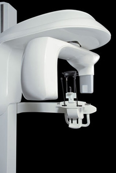 The CS 9600 system gives our oral and maxillofacial practice state-of-the-art technology to help Drs. Herzog, Halkias and Papadimitriou diagnose potential issues more accurately and provide treatment with unprecedented confidence. Unlike a traditional medical CT scanner, our scanner utilizes cone beam CT technology and provides precise, crystal-clear digital images while minimizing your exposure to radiation.
Our cone beam CT scan system enables us to perform a wider range of diagnoses and treatments in our office, helping to reduce multiple visits. The CS 9600 scanner allows us to choose the field of view, or scanning area, that best suits your specific treatment needs. This helps to limit your radiation exposure because we are focusing specifically on your area of concern.
The CS 9600 system brings the latest 3D technology to Oral Surgery Associates, providing unmatched visualization of anatomical detail which aids in treatment planning and helps us to better explain the particulars of your case, as well as address any questions you may have. Drs. Herzog, Halkias and Papadimitriou can use this innovative technology to quickly and easily share 3D images of the area of concern with your referring doctor – allowing the doctors to collaborate on your care, improving your experience, and delivering a positive treatment outcome.
INTRAORAL SCANNER
We utilize an intraoral scanner to obtain digital “impressions” of your upper and lower jaws and teeth. The traditional method of obtaining impressions of your jaws and teeth utilizes a plastic or metal tray filled with semi-liquid impression material that captures the shape of your teeth and jaws. The modern method of obtaining an impression uses digital technology to obtain the same information.

The benefits of intraoral scanning are that it is more comfortable for the patient because it minimizes gagging on the impression material, it is cleaner, and is highly accurate.
We digitally integrate the images obtained with our intraoral scanner with the images obtained with the cone beam CT scanner to help fabricate custom made surgical guides for our more complex and esthetic dental implant cases.
With the use of our cone beam CT scanner and our intraoral scanner we are able to provide the most accurate surgical planning for implant cases to minimize risk of injury to adjacent teeth and vital anatomy as well as ensuring that your implants are placed in the most ideal position for the best esthetic result.